
FIG. 3.
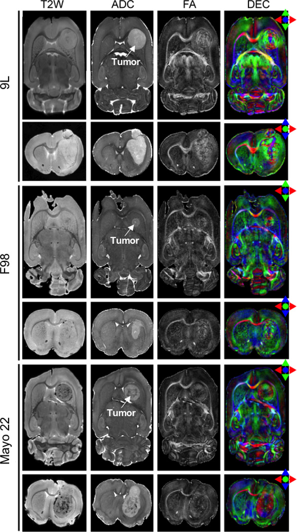
Ex vivo MR images from perfused rat brain samples with 9L, F98, and Mayo 22 glioblastoma xenografts. Post-implantation days were 11 (9L), 10 (F98), and 26 (Mayo 22). Transverse sections and coronal sections are shown. The tumor regions are hyperintense on the ADC images. The contrast between the tumor and surrounding brain tissues on the T2W images is low. All three types of tumors show various degrees of high-level diffusion anisotropy and a unique arrangement of tissue diffusion orientation.