
FIG. 4.
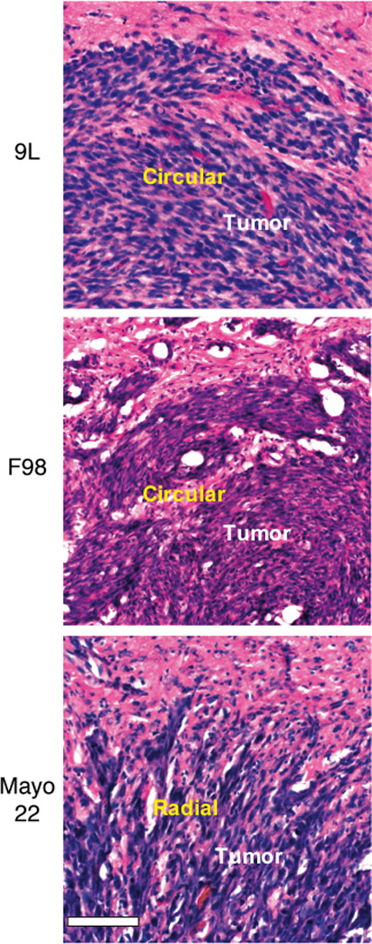
High magnification (20×) histology of 9L, F98, and Mayo 22 tumors. Post-implantation days were 11 (9L), 11 (F98), and 26 (Mayo 22). Bar was 0.1 mm. The morphology of individual tumor cells (blue) and the orientations of interstitial space (white) in the outer parts of the tumors (corresponding to the rims) form a circular pattern (9L and F98) or a radial pattern (Mayo 22). Note the collections of radially organized Mayo 22 tumor cells extending from tumor margins into surrounding brain.