
FIG. 5.
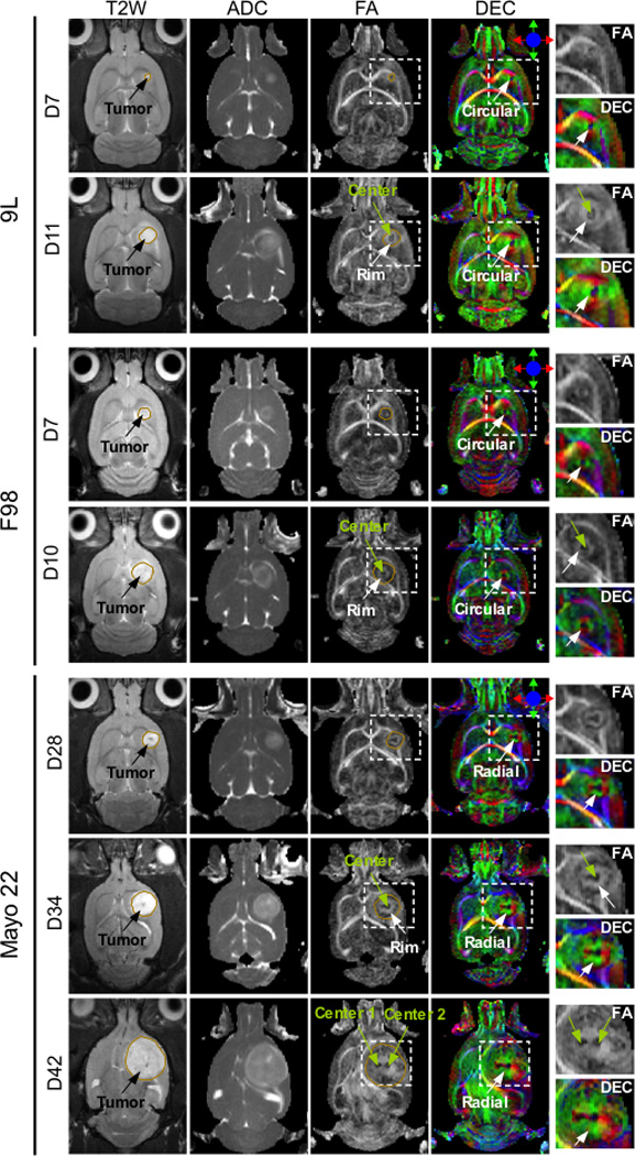
In vivo MR images of 9L, F98, and Mayo 22 xenografts at different stages of tumor growth. A 9L tumor at 7 and 11 days post-implantation, an F98 tumor at 7 and 10 days post-implantation, and a mayo 22 tumor at 28, 34, and 42 days post-implantation. The boundaries of T2W hyperintense tumor regions were manually defined and overlaid on FA images. In the FA and DEC images, tumor regions (inside the rectangular box) were enlarged and shown next to the DEC images. The tumors grow with post-implantation time, as observed within conventional T2W and ADC images. The diffusion patterns appear at an early stage and enlarge with tumor growth in all three models. DTI reveals the presence of two centers for Mayo 22 xenografts, which are not visible with conventional MR images.