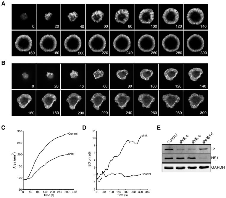
FIGURE 1.
Itk-deficient T cells exhibit unstable lamellipodial protrusions. A, Jurkat T cells stably expressing GFP-actin were transfected with empty suppression vector. After 72 h, cells were plated on coverslips coated with anti-CD3 and imaged by confocal microscopy for the indicated times (seconds). Selected images from one time-lapse series are shown; these correspond to supplemental Video 1. B, Jurkat cells stably expressing GFP-actin were transfected with Itk suppression vector and analyzed as in A. Selected images from one time-lapse series are shown; these correspond to supplemental Video 3. C, The contact area of each cell at each time point was determined, and the average was calculated for each cell population at each 5-s time point (control = 46 cells; shItk = 49 cells). D, Irregularity of cell shape was assessed by measuring the radial variance for each cell at each time point and calculating the average values. E, Western blot analysis showing suppression of Itk or HS1 in GFP-actin Jurkat cells transfected with the indicated suppression vectors.

