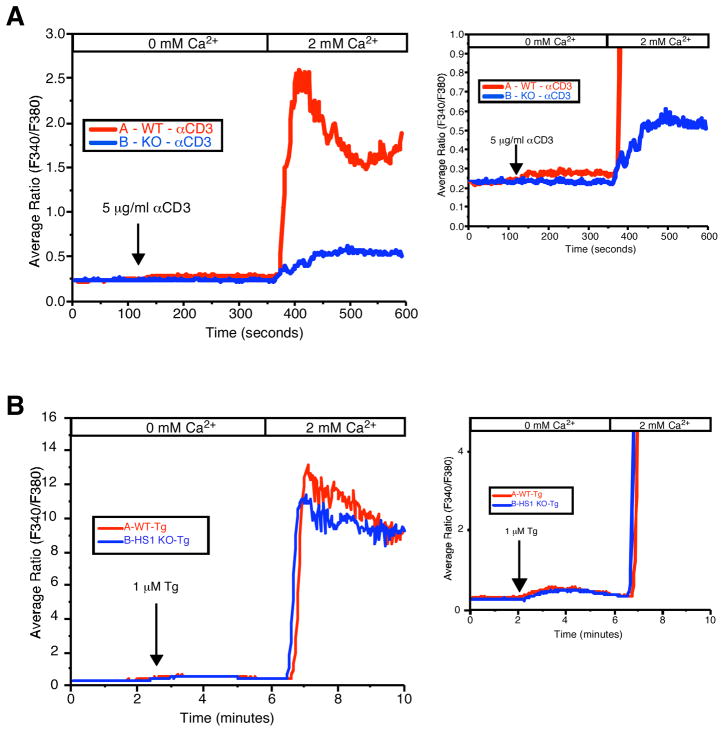
FIGURE 5.
HS1 is required for release of Ca2+ from intracellular stores. A, CD4+ T cells from HS1+/+ or HS1−/− mice were loaded with the fura 2-AM and plated on poly(L-lysine)-coated coverslips. Immediately before TCR stimulation, the cells were superfused with Ca2+-free bath solution. At the indicated time, cells were stimulated by the addition of anti-CD3 (500.A2) and analyzed by fluorescence microscopy to visualize a rise in cytoplasmic Ca2+ indicative of release from ER stores. Extracellular Ca2+ was then added to visualize Ca2+ influx. Right, An expansion of the y-axis to visualize release from ER stores. B, Cells prepared as in A were treated with 1 μM thapsigargin (Tg) and imaged as in A.

