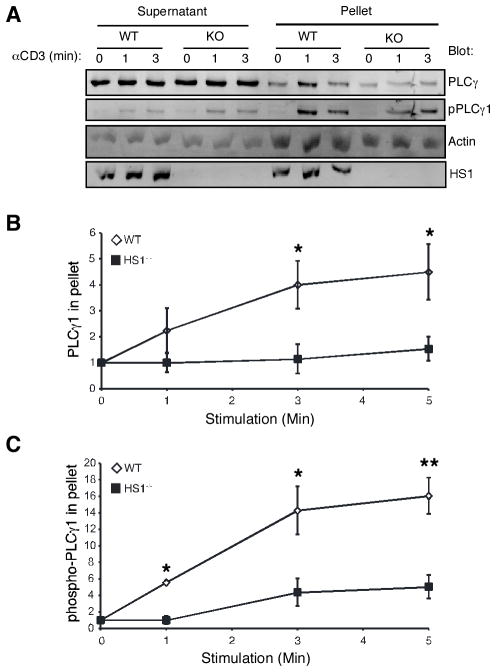
FIGURE 7.
HS1 is required for PLCγ1 cytoskeletal association. A, T cells from HS1+/+ or HS1−/− mice were stimulated as indicated, lysed in cytoskeletal stabilizing lysis buffer, and separated into cytosol-enriched supernatant and F-actin-rich pellet fractions. Fractions were analyzed by Western blotting, as indicated. pPLCγ1 was detected using an Ab specific for PLCγ1 phosphorylated at Y783. B, The amount of PLCγ1 in the pellet fractions at each time point was quantified and normalized to the amount of PLCγ1 in the pellet in unstimulated cells. Data represent averages from four independent experiments ± SEM. *, p < 0.05. C, The amount of PLCγ1 phosphorylated at Y783 in the pellet fractions at each time point was quantified and normalized to the amount of PLCγ1 phosphorylated at Y783 in the pellet in unstimulated cells. Data represent averages from four independent experiments ± SEM. *, p < 0.05; **, p < 0.01.

