
Figure 3.
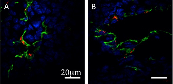
A, B: Representative confocal fluorescent images of the tumor endothelium (anti-CD31 stain-ing, green), β3-integrin (anti-CD61 staining, red), and nuclei (DAPI, blue). The expression of β3-integrin was co-localized with tumor endothelium. The scale bar is 20 μm.