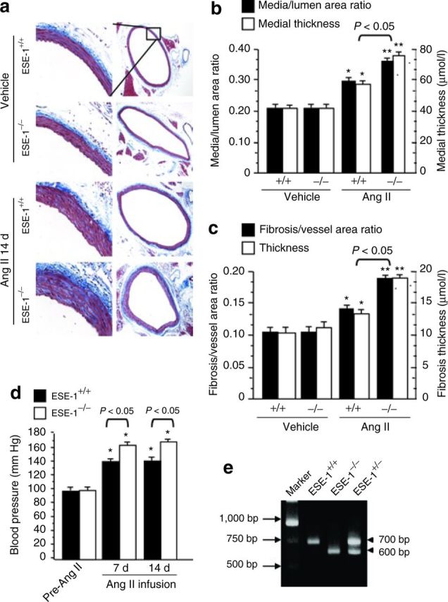
Figure 3. Increase in Ang II–induced vascular remodeling and high blood pressure in ESE-1−/− vs. ESE-1+/+ mice. (a) Comparison of the effect of Ang II infusion vs. vehicle controls on perivascular fibrosis and arterial thickening in the aorta of ESE-1+/+ vs. ESE-1−/− mice. Aortic sections were stained with Masson's trichrome stain. Original magnification on left panel ×250 and right panel ×20. (b,c) Statistical analysis of aortic thickness (b) and perivascular fibrosis (c) in ESE-1+/+ vs. ESE-1−/− mice compared to vehicle controls. *P < 0.05 vs. vehicle of mice. **P < 0.01 vs. vehicle of mice (n = 5 per group). (d) The systolic blood pressure of mice was measured by using a Visitech BP-2000 system (see Methods) before (pre-Ang II) and after Ang II infusion (1.4mg/kg/day) for 1 week (7 days) and 2 weeks (14 days). *P < 0.01 vs. pre-Ang II of mice (n = 5 per group). (e) Representative PCR result for genotype of the ESE-1+/+, ESE-1+/−, and ESE-1−/−. Ang II, angiotensin II; ESE-1, epithelium-specific ETS transcription factor-1; PCR polymerase chain reaction.

