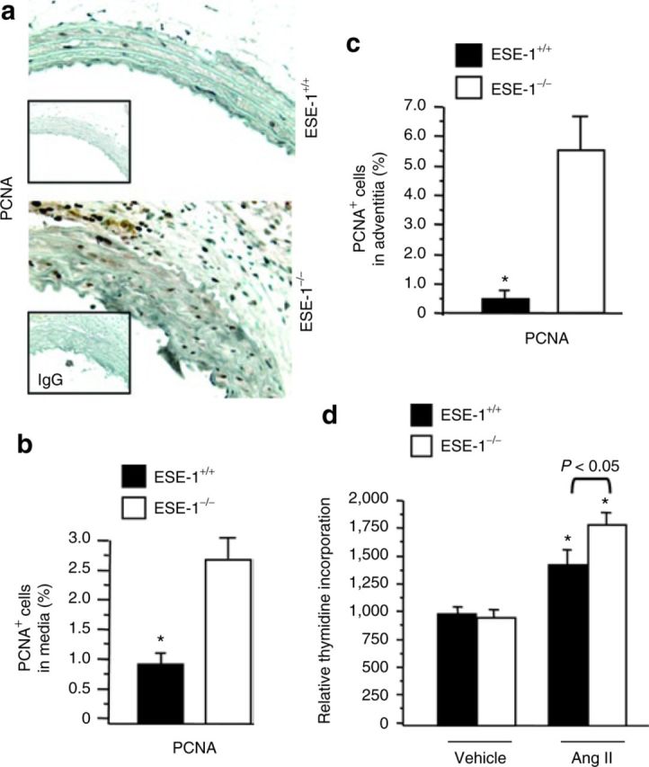
Figure 4. Immunohistochemical staining of PCNA in the thoracic aorta of ESE-1+/+ and ESE-1−/− mice after Ang II infusion. (a) Evaluation of cell proliferation by PCNA staining after 2 weeks of Ang II infusion (1.4mg/kg/day) in the thoracic aorta of ESE-1+/+ compared to ESE-1−/− mice. Original magnification ×250. Isotype-matched controls are shown below each panel. (b,c) Statistical analysis of the positive cell staining with PCNA in media (b) and in adventitia (c). (d) Effect of Ang II on [3H]thymidine uptake. Quiescent human aortic smooth muscle cells were stimulated with Ang II (100nmol/l) for 18h and pulsed with [3H]thymidine for 5h. Incorporation of [3H]thymidine was measured by liquid scintillation spectrophotometer (n = 3). Values represent mean ± s.e.m. *P < 0.01 vs. vehicle, respectively. PCNA, proliferating cell nuclear antigen. Ang II, angiotensin II; ESE-1, epithelium-specific ETS transcription factor-1; IgG, immunoglobulin G.

