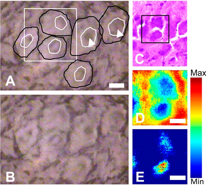
Figure 1.

SR-FTIR-FPA imaging of pyramidal neurons within the hippocampus. (A) Optical image of the unstained tissue section with locations of neuron soma and nuclei drawn free hand in black and white, respectively. White square indicates region analyzed with SR-FTIR-FPA imaging. (B) Optical image of (A) without annotations. (C) Optical image of H&E stained section analyzed with SR-FTIR-FPA imaging (black box defines region analyzed). H&E stain performed after spectroscopic analysis. (D) False-color functional group image generated from area underneath the lipid ν(C=O) band (1750–1710 cm–1), to reveal the presence of neuron soma. (E) False-color functional group image generated from the ratio of the area underneath the νs(CH3) band (2880–2865 cm–1) and νs(CH2) band (2860–2845 cm–1), to reveal the presence of neuron nuclei. Scale bars = 10 μm.
