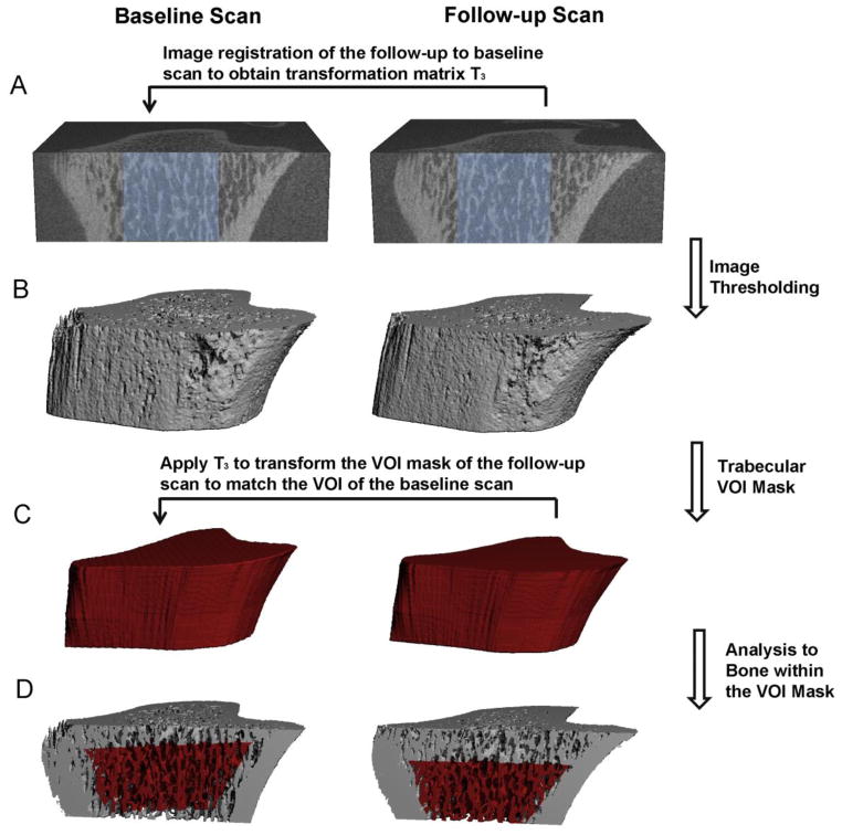
Figure 2.
Schematics of the image registration and microstructural analysis of trabecular bone: (A) Image registration was performed on greyscale images of a trabecular subregion (shown in light blue) to obtain the translation matrix T3 to transform follow-up scan to the baseline scan. (B) Image thresholding was performed to distinguish bone matrix from bone marrow and background. (C) A trabecular VOI mask was generated based on the thresholded follow-up scan to separate trabecular from cortical compartment. Then the T3 was applied to transform the VOI mask of the follow-up scan to the corresponding VOI of the baseline scan. (D) Trabecular microstructural analysis was then performed on the trabecular bone within the correponding VOIs (highlighted red region) of both baseline and follow-up scans.

