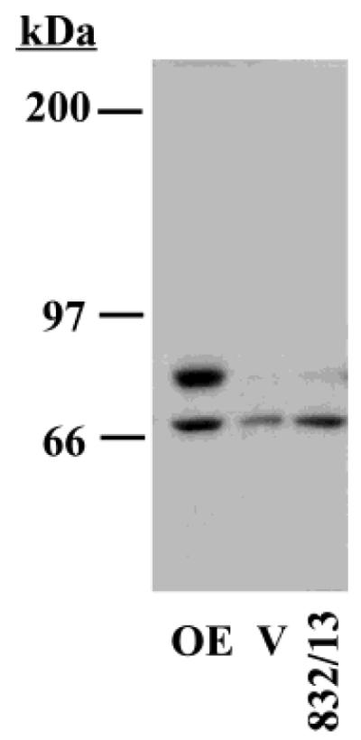
Figure 1.

Immunoblotting analyses of iPLA2β-immunoreactive protein expression in 832/13 INS-1 cells. Aliquots (50 μg) of cytosol prepared from INS-1 cells transfected with either an empty vector (V) or vector containing iPLA2β cDNA construct (OE) and from 832/13 INS-1 cells were analyzed by SDS–PAGE, and the proteins were transferred onto immobolin-P PVDF membrane. The electroblot was then processed for immunoblotting analyses, and immunoreactive iPLA2β protein was visualized by ECL.
