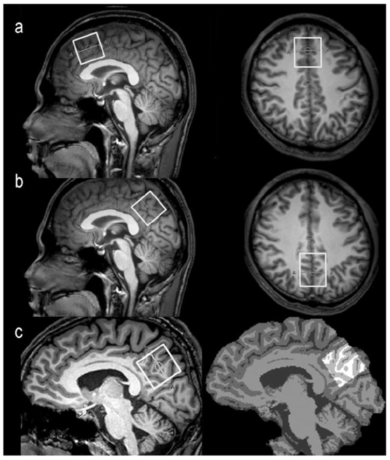
Fig.1.

The VOIs position in the frontal region (a) and parietal region (b) for spectroscopic measurement using a MEGA-PRESS sequence. The white box represents the location of the VOI (3 × 3 × 3 cm3) in the sagittal and axial images. (c) The 3D T1-weighted brain images (left) were segmented as gray matter (GM), white matter (WM), or cerebrospinal fluid (CSF) using the FAST (FMRIB’s automated segmentation tool) of the “FSL” software package (right) and the VOIs were re-created using the “Re-creation of VOI” Matlab tool.
