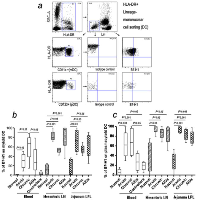
Figure 1.
(a) Rhesus macaque myeloid DC and plasmacytoid DC were identified in blood as HLA-DR+, lineage negative (Lin-) cells as depicted. PBMCs were first selected based on side scatter and HLA-DR+ (R1 gate) and further defined as HLA-DR+ Lin- cells (R2 gate). CD11c+HLA-DR+ (mDCs) and CD123+ HLA-DR+ (pDCs) events within R2 were defined in R3 and R4, respectively. B7-H1 expression on macaque mDCs or pDCs in blood is shown in representative histograms. (b and c) Expression of B7-H1 on myeloid DC and plasmacytoid DC from blood and mucosal tissues in SIV-infected macaques. Note marked and significant increases in B7-H1 expression in blood, mes LN, and jejunum pDCs and mDCs in SIV infected animals in acute and chronic infection. Mean values, SE, and statistically significant differences are shown.

