Figure 3.
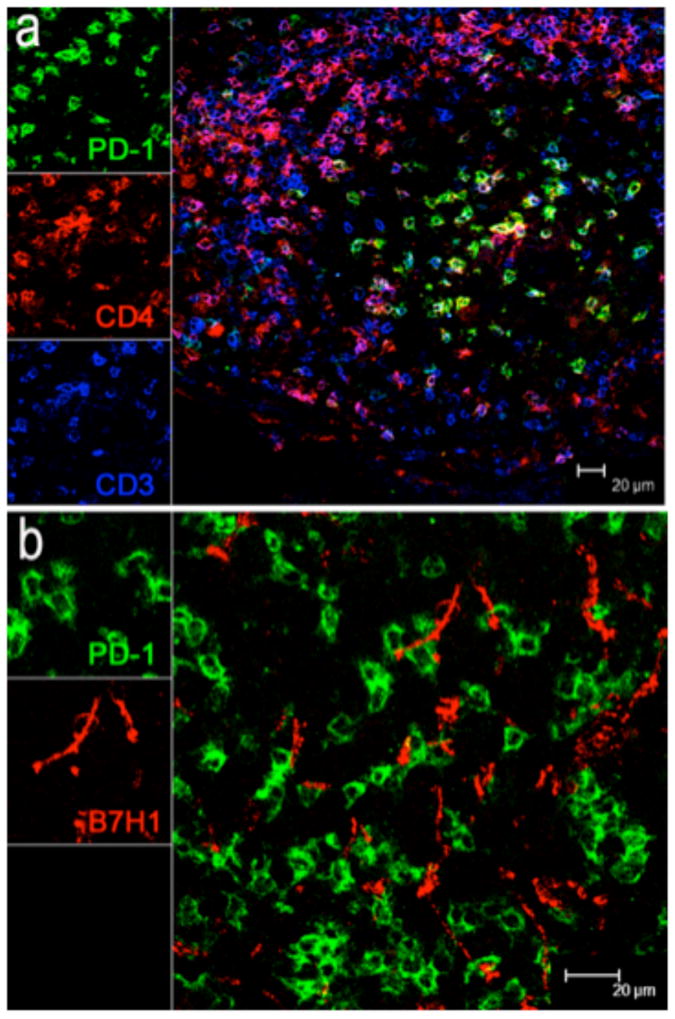
(a) Distribution and co-expression of PD-1 on CD4+ T (CD3+) cells in lymph nodes of a representative SIV-infected macaque by confocal microscopy. Immunohistochemistry demonstrates PD-1 (green) expression is primarily co-expressed on CD4+ T cells in germinal centers in chronic SIV infection. (scale bar = 20μm). (b) Co-localization of B7-H1 (red) and PD-1 (green) expression in lymph node of a chronically infected macaque 3 months after infection. Note B7-H1+ cells (red) have a dendritic morphology and co-localize with PD-1+ cells (green) which have a lymphocyte morphology and shown to be CD4+ T cells in (a). B7-H1+ cells (red) are mostly found in follicular areas, and usually in close proximity and often direct contact with PD-1+CD4+ T cells. (scale bar = 20μm).

