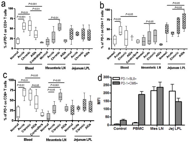
Figure 4.
PD-1 expression on T cells in blood, mesenteric LN and lamina propria in SIV-infected macaques. (a, b and c) Percentage of PD-1 expression on CD3+ (a), CD4+ (b) and CD8+ T cells (c) in normal, acute, chronic, and AIDS stage infection, and symptomatic AIDS. Note marked expansion of PD-1+ T cells occurs and persists in most tissues throughout SIV infection. (d) Surface PD-1 is predominantly expressed on SIV-specific CD8+ T cells clone (CM9 and SL-8) from chronic SIV-infected macaques. Mean values, SE, and statistically significant difference are shown.

