
Figure 7.
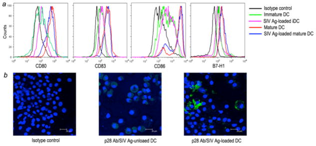
Identification of mature DCs and SIV antigen-loaded DCs by flow cytometry (top) and fluorescent microscopy (bottom). (a) Histogram of mature monocyte-derived macaque DCs. Monocyte-derived immature DCs (iDC) were cultured with maturation cocktail and phenotyped by FACS analysis of FITC-anti-CD80 or -CD86, PE-anti-CD83 or -B7-H1, and PerCP-anti-HLA-DR. (b) Immunofluorescent staining of SIV lysate pulsed DCs. Immature DCs were pulsed with SIV lysate for 24 hours, and then incubated for 2 days in DC maturation cocktail. SIV lysate-unloaded and -loaded DCs were cytospun onto slides, fixed and stained with a control antibody (left), or anti-p28 monoclonal antibody (green). Nuclei are stained in blue. Representative fields (scale bar = 20μm) SIV p28 antigen staining of loaded and unloaded DCs are shown.