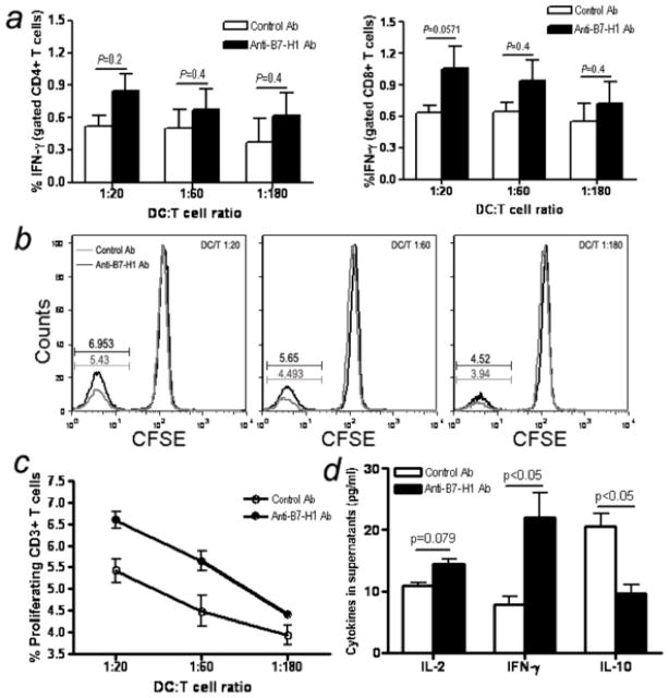
Figure 8.
Effects of B7-H1 blockade on SIV-specific antigen function. (a) Loaded and unloaded DCs were added to 5×10⁵ autologous T cells at shown ratios with control or anti-B7-H1 Ab added at dilutions indicated. Intracellular IFN-γ staining was performed 6 hours after incubation. Means ±SEM of triplicate data are shown. (b and c) Effects of B7-H1 blockade on SIV-specific T cell proliferation. Ranging doses of the loaded DCs were added to 105 autologous T cells, with control or anti-B7-H1 Ab added as indicated and T cell proliferation was assessed by CFSE dilution 6 days later. b, histogram of CFSE dilution. Means (±SEM) of triplicate data are provided. DCs and T cells alone (±SIV lysates) served as negative controls for both assays, which showed background levels less than the unloaded DC-T cells controls. (d) IL-2, IFN-γ and IL-10 cytokine levels in supernatants of T/DC co-culture at 60:1 after 6 days incubation. Bars reflect means of three independent data points.

