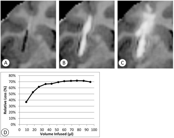
Figure 3.
Extreme example of backflow loss (Cy0140-left). Coronal T1-maps (negated) centered at the catheter position illustrate that backflow along the catheter shaft can be a major cause of infusate loss out of the putamen. In this extreme example, about 70% of the infusate flows outside of the nonhuman primate putamen. A: Shows the pre-infusion catheter position. B: backflow is clearly developed outside the putamen, 13 mm from the catheter tip, at 18 minutes. C: Most of the infusate flows out the top of the putamen and spreads through the frontal white matter tracts above the putamen as the infusion continues.

