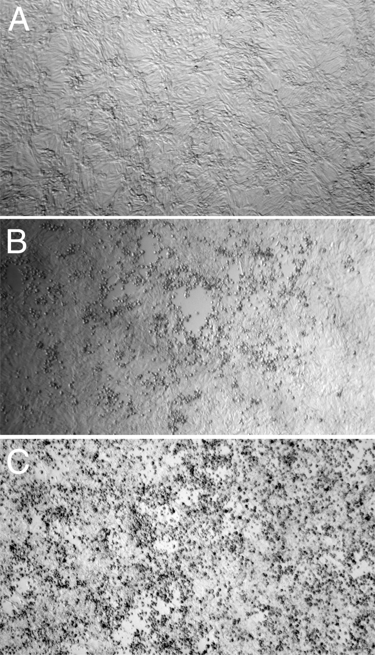
Fig 1.
Cytopathic effect of the CVV-infected Vero cells. Vero cells were seeded in a 24-well plate and grown to 90% confluence and were either mock infected (A) or infected at a multiplicity of infection (MOI) of 0.3 (B) or at an MOI of 30 (C) with the MNZ-92011 strain, which was harvested from BGMK cells, amplified, and titrated in Vero cells. Photographs were taken on day 4 postinfection, using a Spot RT-KE monochrome camera connected to a Nikon Diaphot TMD microscope at ×40 magnification and Spot 5.0 Basic software.

