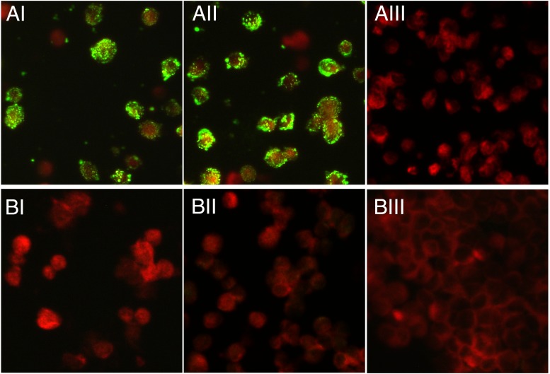
Fig 2.
Detection of anti-CVV antibodies in the patient's convalescent-phase serum. The results of an IFA of the patient's 24-day convalescent-phase serum (I), an anti-CVV mouse hyperimmune serum (II), and an anti-La Crosse virus human convalescent-phase serum (III), using Caco2 cells that were mock infected (BI to BIII) or at day 2 postinfection with the MNZ-92011 strain (AI to AIII), are shown. Fluorescein isothiocyanate (FITC)-conjugated dual anti-human/anti-mouse IgG goat antibody (Focus Diagnostics, Cypress, CA) was used as a secondary antibody. Images were taken with a Zeiss AxioCam MRc camera attached to AxioImager A1 microscope at ×400 magnification, using Zeiss AxioVision 3.1 software. All sera were at a dilution of 1:16, with the exception of the anti-CVV mouse serum represented in panels AII and BII, where the serum was diluted 1:1,024.

