
Fig 5.
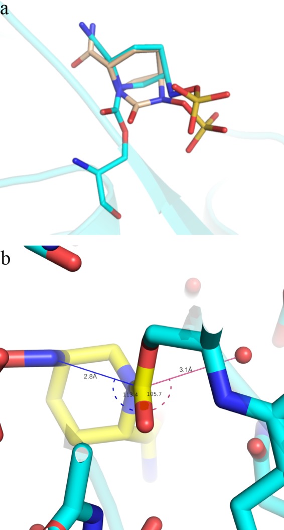
Substrate-like conformation of avibactam in the enzyme pocket. (a) Overlay of covalently modified avibactam on CTX-M-15 (cyan) on a closed avibactam model (light brown). The protein backbone is shown in cartoon form. (b) Bond distance and angle near recyclization event in CTX-M-15. Avibactam is depicted as a yellow stick, while Ser70 is in cyan. The distance between deacylating water (shown as red spheres) and carbonyl C7 is shown as a pink line, while that between attacking N6 and carbonyl C7 is a blue line. The angles are depicted in the same color with dashed lines.