
Fig 3.
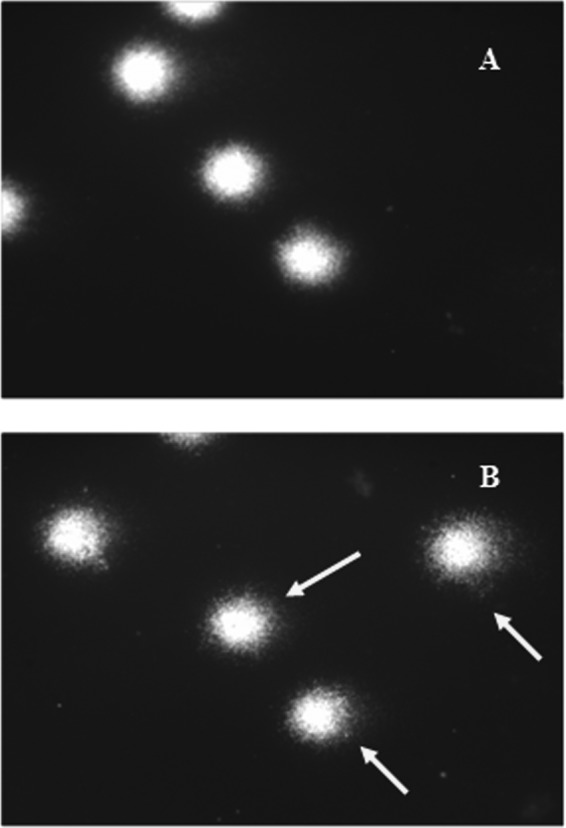
Typical fluorescence microscopy images (comets) of human PBLs left unexposed (A) or exposed to colistin at 1,100 ng/ml (B). The arrows show comet tails indicating DNA damage of leukocytes after treatment with colistin.
Official websites use .gov
A
.gov website belongs to an official
government organization in the United States.
Secure .gov websites use HTTPS
A lock (
) or https:// means you've safely
connected to the .gov website. Share sensitive
information only on official, secure websites.
Typical fluorescence microscopy images (comets) of human PBLs left unexposed (A) or exposed to colistin at 1,100 ng/ml (B). The arrows show comet tails indicating DNA damage of leukocytes after treatment with colistin.