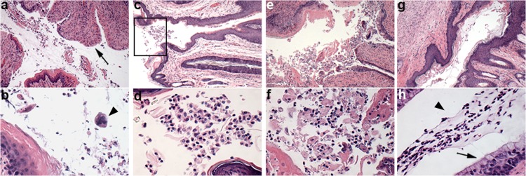
Fig 8.
Representative photomicrography of vaginal tissue sections. (a, b) Group 1 (infected, untreated) showing denuded epithelium (arrow) and multinucleated cell (arrowhead); (c, d) group 7 (infected, MI-S pretreatment), vaginal orifice (boxed); (e, f) group 8 (infected, vehicle pretreatment); (g, h) group 3 (uninfected, MI-S treatment) showing mucified columnar epithelium (arrow) and mucus with epithelial cells in the lumen (arrowhead). Slides were stained with H&E; magnification for first and second rows, ×100 and ×400, respectively.

