
Fig 3.
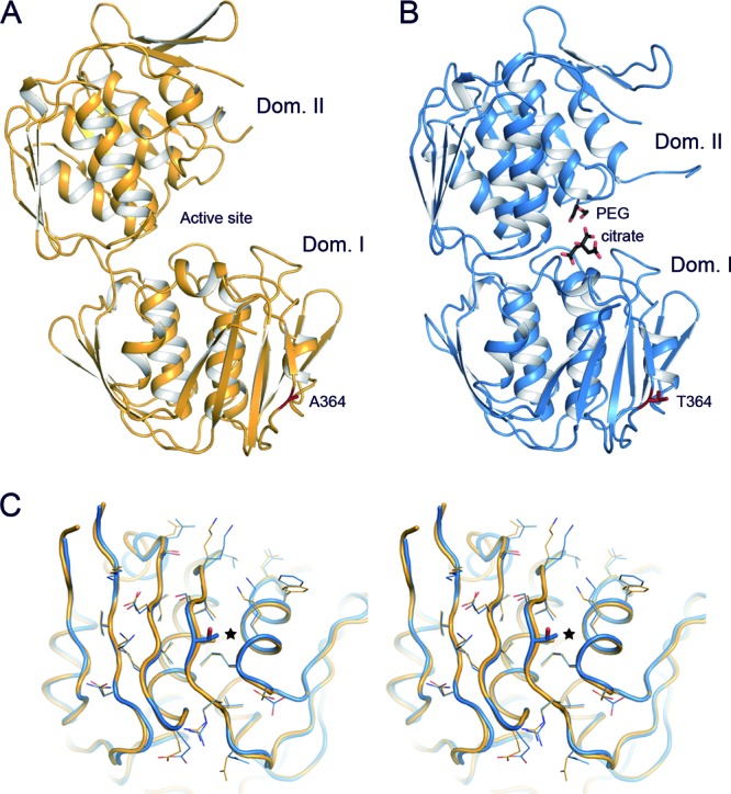
Crystal structure of pneumococcal MurA1. (A) Overall structure of S. pneumoniae MurA1D39 in an open conformation. Ala364 is represented as capped sticks and colored red. (B) Overall structure of S. pneumoniae MurA1Hungary19A in a closed conformation, showing a citrate and a PEG molecule in the catalytic pocket (black-capped sticks). Thr364 is represented as capped sticks and colored red. (C) Stereo view of the superposition of domain I of MurA1D39 (orange) and MurA1Hungary19A (blue). The Ala364Thr mutation is represented in sticks and highlighted (star). Side chains of residues around position 364 are drawn as lines to show that there are no relevant modifications upon mutation. Dom., domain.