
Fig 1.
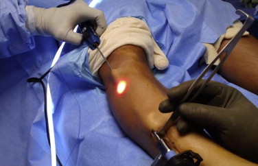
Clinical photograph of the right leg of a patient in the supine position. A 30° arthroscope is placed through a small distal incision. The Spectrum suture passer is then placed through a proximal incision to introduce a No. 1 PDS. The technique used for the anterior compartment is depicted.