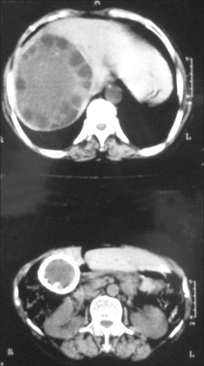
Figure 1.

CECT abdomen: Documented hydatid cyst in right lobe of the liver. Another cystic lesion with thick calcified wall was seen in segment-VI of liver measuring 5×5 cm. A calcified area 2 cm in size was also seen medial to the above-calcified cystic lesion. The gall bladder was not seen separately from the calcified cystic lesion. IHBR and CBD were normal
