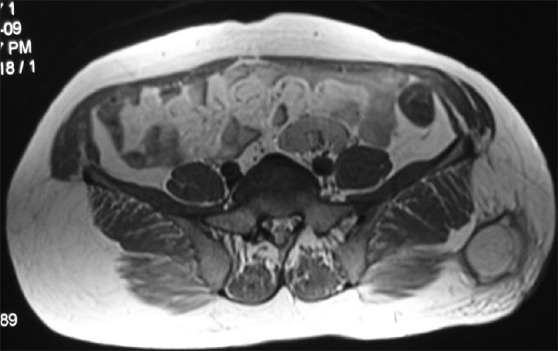
Figure 3.

MRI showing multicystic swelling overlying gluteus maximus which is hyperintence on T2WI/STIR and hypointence on T1WI images. Thick septa seen within and surrounding tissue showing edema

MRI showing multicystic swelling overlying gluteus maximus which is hyperintence on T2WI/STIR and hypointence on T1WI images. Thick septa seen within and surrounding tissue showing edema