Figure 4.

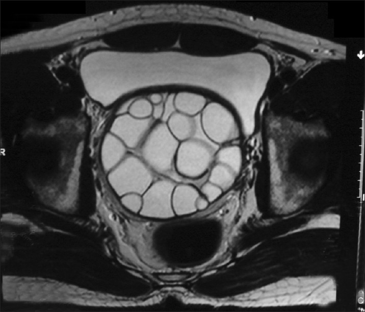
Axial T2WI (MRI) showing hyper intense, multicystic lesion with multiple daughter cysts in relation to the right seminal vesicle

Axial T2WI (MRI) showing hyper intense, multicystic lesion with multiple daughter cysts in relation to the right seminal vesicle