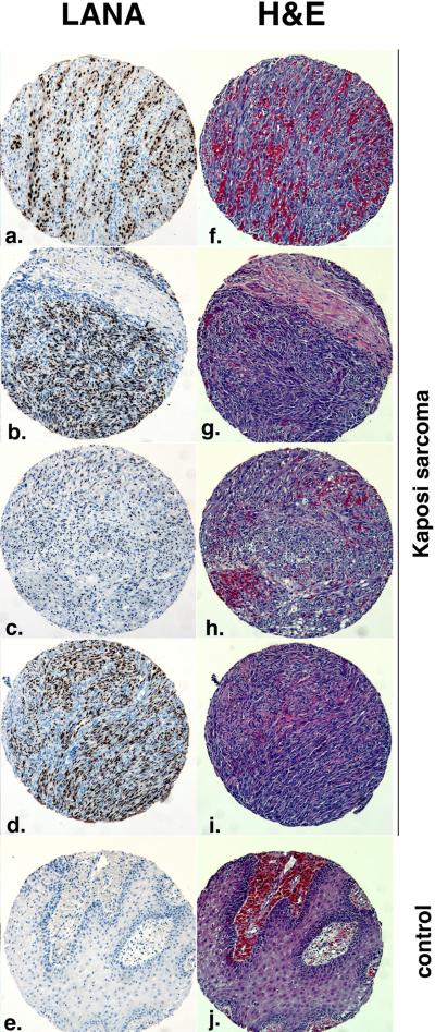
Figure 2.
Different histological examples of KS. Shown is immunohistochemistry for the KSHV LANA protein in brown (panels a to d) and Hematoxilin & Eosin (H&E) stain of lesions (panel f. – i.) . Panels e and j represent non-KS control. The samples were part of the first KS tissue microarray made available through the AIDS Cancer Specimen Resource (ACSR). Magnification 40x.

