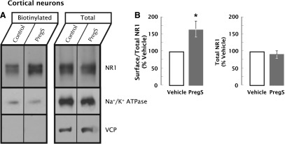
Fig. 9.

PregS increases surface NR1 subunits on neocortical neurons. (A) Representative immunoblot of surface NR1 levels in neocortical neurons exposed to PregS for 10 minutes as compared with vehicle. Surface biotinylated proteins were affinity purified and analyzed with a fraction of total cellular proteins by immunoblotting with antisera to NR1, surface Na+/K+ ATPase (positive control), or cytoplasmic valosin-containing protein (VCP) (negative control). (B) Quantitation of the effect of PregS (50 μM, 0.1% DMSO, 10 minutes) on total and surface-biotinylated NR1. (Left panel) Immunoreactivity in biotinylated aliquots as a fraction of total NR1 expressed as a percentage of vehicle. PregS induced a 62% ± 25% increase (*P = 0.033, n = 6 experiments, Student’s t test) in the biotinylated fraction of NR1. (Right panel) There was no change in total NR1 levels with PregS treatment (P = 0.291, n = 6 experiments, Student’s t test).
