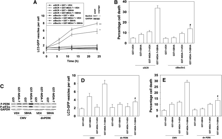
Fig. 2.
Induction of ER stress and autophagy plays a role in the interaction between MDA-7/IL-24 and HDACIs. (A) GBM6 cells were transfected in quadruplicate with a plasmid to express an LC3 (ATG8)–GFP fusion protein and in parallel transfected with scrambled small interfering RNA (siRNA; siSCR) or an siRNA to knock down Beclin1 expression. Twenty-four hours after transfection, the cells were treated with GST or GST-MDA-7 (20 nM) and/or SBHA (3 μM). Cells (a representative of 40 per well per time point) were examined 6, 12, and 24 hours after treatment using an Axiovert microscope (40×) for the formation of punctate vesicles containing LC3-GFP. Data are plotted as the number of LC3-GFP vesicles per cell (n = 2, ± S.E.M.). *P < 0.05 greater than GST + VEH (vehicle); **P < 0.05 greater than GST-MDA-7 + VEH (vehicle). (B) GBM6 cells were transfected with siSCR or an siRNA to knock down Beclin1 expression. Twenty-four hours after transfection, the cells were treated with GST or GST-MDA-7 (20 nM) and/or SBHA (3 μM). Cells were isolated 48 hours later, and viability was determined by trypan blue exclusion (n = 3, ± S.E.M.). #P < 0.05 less than corresponding value in siSCR cells. (C) GBM6 cells were transfected with an empty vector control plasmid or a plasmid to express dominant negative (dn) PERK. Twenty-four hours after transfection, the cells were treated with GST-MDA-7 (20 nM), SBHA (3 μM), or the agents combined. Cells were isolated 6 hours after exposure, and the phosphorylation of PERK and eIF2α determined (representative n = 2). (D) GBM6 cells were transfected in quadruplicate with an LC3 (ATG8)–GFP fusion protein and in parallel transfected with an empty vector control plasmid or a plasmid to express dominant negative PERK. Twenty-four hours after transfection, the cells were treated with GST or GST-MDA-7 (20 nM) and/or SBHA (3 μM). Cells (a representative of 40 per well per time point) were examined 24 hours after treatment using an Axiovert microscope (40×) for the formation of punctate vesicles containing LC3-GFP. Data are plotted as the number of LC3-GFP vesicles per cell (n = 3, ± S.E.M.). #P < 0.05 less than corresponding value in CMV cells. (E) GBM6 cells were transfected with an empty vector control plasmid or a plasmid to express dominant negative PERK. Twenty-four hours after transfection, the cells were treated with GST-MDA-7 (20 nM), SBHA (3 μM), or the agents combined. Cells were isolated 48 hours later, and viability was determined by trypan blue exclusion (n = 3, ± S.E.M.). #P < 0.05 less than corresponding value in CMV cells.

