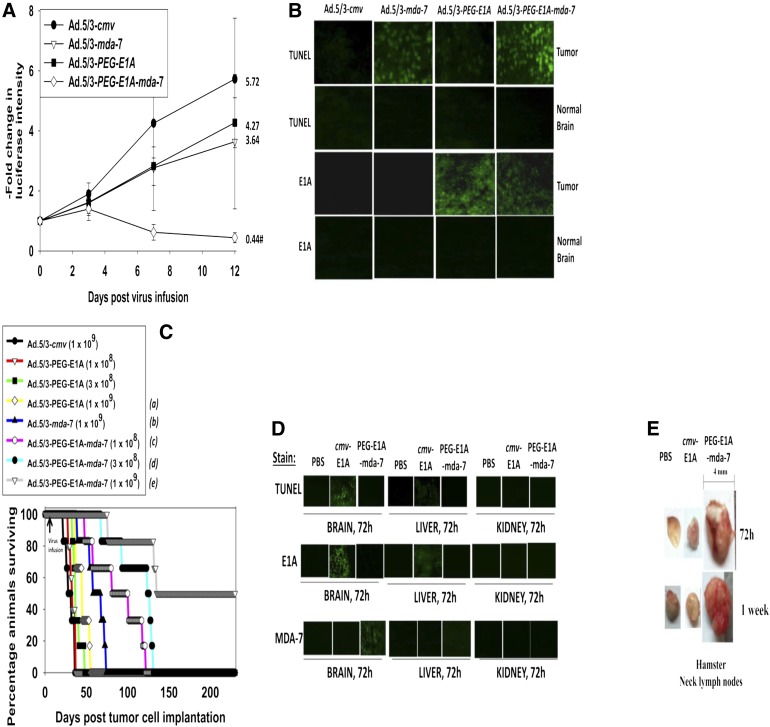
Fig. 7.
Ad.5/3-PEG-E1A-mda-7 (Ad.5/3-CTV) prolongs animal survival in a dose-dependent fashion and does so to a greater extent than Ad.5/3-mda-7. (A) GBM6-luciferase cells (0.5 × 10⁶) were implanted into the brains of athymic mice. Seven days later, the tumors were infused with 1 × 10⁸ pfu of Ad.5/3-cmv, Ad.5/3-mda-7, Ad.5/3-PEG-E1A, or Ad.5/3-PEG-E1A-mda-7 (Ad.5/3-CTV). On the days indicated in the graph, the animals were injected with luciferin (150 mg/kg) and were imaged 15 minutes later after placement into an IVIS Xenogen imager (PerkinElmer, Waltham, MA). The fold increase in luciferase intensity for the mean of each animal group was plotted (n = 2, six animals total ± S.E.M.). #P < 0.05 less than Ad.5/3-cmv, Ad.5/3-mda-7, or Ad.5/3-PEG-E1A (Ad.5/3-CTV). The fold change in luciferase activity at day 12 is shown numerically. (B) Brains from animals at day 12 (panel A) were removed, fixed in OCT compound (Tissue Tek), and cryostat sectioned (Leica) as 12 μm sections. Sections from tumor tissue and normal brain were stained for apoptosis (TUNEL) and for expression of the viral E1A protein. (C) GBM6 cells (0.5 × 10⁶) were implanted into the brains of athymic mice. Seven days later, the tumors were infused with Ad.5/3-cmv (1 × 10⁹ pfu), Ad.5/3-mda-7 (1 × 10⁹ pfu), Ad.5/3-PEG-E1A (1 × 10⁸; 3 × 10⁸; 1 × 10⁹ pfu), and Ad.5/3-PEG-E1A-mda-7 (Ad.5/3-CTV) (1 × 10⁸; 3 × 10⁸; 1 × 10⁹ pfu). Animal survival was monitored on a daily basis (n = 2, six animals total). aP < 0.04 greater survival than Ad.5/3-cmv; bP < 0.0008 greater survival than Ad.5/3-cmv; cP < 0.04 greater survival than Ad.5/3-mda-7; dP < 0.004 greater survival than Ad.5/3-mda-7; eP < 0.0008 greater survival than Ad.5/3-PEG-E1A-mda-7 (Ad.5/3-CTV) at a dose of 3 × 10⁸ pfu). (D) Syrian hamster brains were infused with PBS, Ad.5/3-cmv-E1A (2 × 10⁹ pfu), or Ad.5/3-PEG-E1A-mda-7 (Ad.5/3-CTV) (2 × 10⁹ pfu). Seventy-two hours after infusion, the animal brains, livers, and kidneys were isolated and fixed. Sections (12 μm) were taken and stained for apoptosis (TUNEL), the levels of viral E1A protein, and the levels of MDA-7/IL-24 protein. (E) Syrian hamster brains were infused with PBS, Ad.5/3-cmv-E1A (2 × 10⁹ pfu), or Ad.5/3-PEG-E1A-mda-7 (Ad.5/3-CTV) (2 × 10⁹ pfu). Seventy-two hours and 1 week after infusion, the animals were sacrificed, and their neck lymph nodes were dissected.

