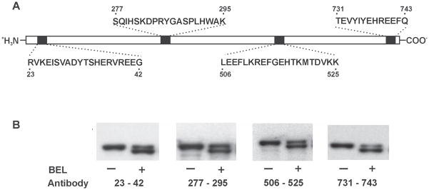
Figure 2. Western blot analysis of BEL-treated iPLA2β utilizing antibodies directed against four different regions of the iPLA2β protein.

Epitopes for the four antibodies are indicated in Panel A. iPLA2β was incubated with (S)-BEL at 22°C for 3 min. Following separation on a 7% SDS-PAGE gel, the protein bands were transferred to Immobilon-P PVDF membranes and probed separately with the four different antibodies against iPLA2β in Panel B. Both native and BEL-treated iPLA2β are immunoreactive to all four antibodies. “−“ indicates the control sample and “+” indicates the BEL-treated sample.
