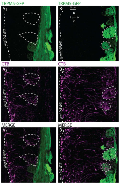
Figure 4.
Confocal images of CTB(+), TRPM5(+) and CTB(+) & TRPM5(+) glomeruli in the MOB. Sections are oriented in the coronal plane. A and B represent example regions of the MOB from two different successfully injected cases with TRPM5-GFP(+) glomeruli in green and CTB(+) mitral cells terminating in the glomerular layer in magenta. A1–3 show an example in which the TRPM5-GFP(+) expression and CTB(+) label do not overlap; dashed polygons encircle glomeruli that are labeled with CTB(+) only. B1–3 highlight examples of TRPM5-GFP(+) expression and CTB(+) label overlap in the glomerular layer; dashed polygons encircle glomeruli that express CTB(+) & TRPM5-GFP(+).

