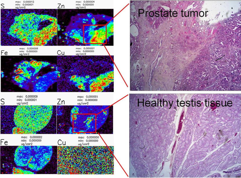
Figure 1.

X-ray fluorescence microscopy of dog testis and prostate tissues. XFM analysis can be performed on sections of paraffin embedded dog tissues to show the distributions of various elements. In this example, both testis and prostate samples were sectioned at 5 μm and stained with H & E. The corresponding x-ray Fluorescence (XFM) scan shows the distribution of sulfur (S), zinc (Zn), iron (Fe) and copper (Cu) in both tissues.
