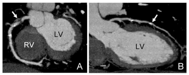
Figure 1.

Coronary computed tomography of 56 year-old male with acute coronary syndrome and a history of cocaine use demonstrating non-calcified, calcified plaque, and a significant coronary stenosis. A: Maximum intense projection (MIP) image of the right coronary artery with non-calcified plaque in the proximal portion (arrow) and a significant coronary stenosis (>50% luminal narrowing) in the proximal segment of the vessel. B: Curved multiplanar reformation of the left anterior descending coronary artery with calcified plaque in the mid segment of the vessel. LV: left ventricle; RV: right ventricle.
