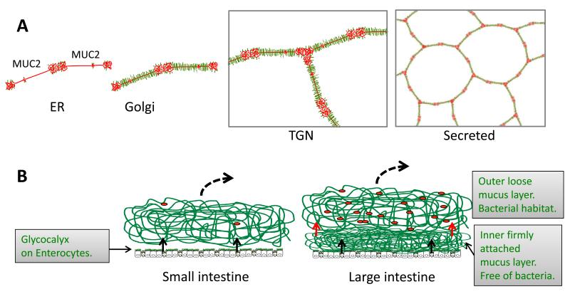
Fig.1.
Schematic outline of the MUC2 mucin and its formation of mucus in the small and large intestine. A. Assembly of the MUC2 mucin (protein core red) into dimeric forms in the endoplasmic reticulum (ER), O-glycosylation (green) in the Golgi apparatus, formation of trimeric forms in the trans Golgi network (TGN) and a schematic picture of the secreted MUC2 polymer. B. The MUC2 mucin is secreted from the goblet cells (black arrows) to form the mucus. Colon have a two layered mucus where the inner layer is converted (red arrows) to the outer mucus layer. The stomach has a mucus essentially as that of colon (not shown). Red dots symbolizes bacteria.

