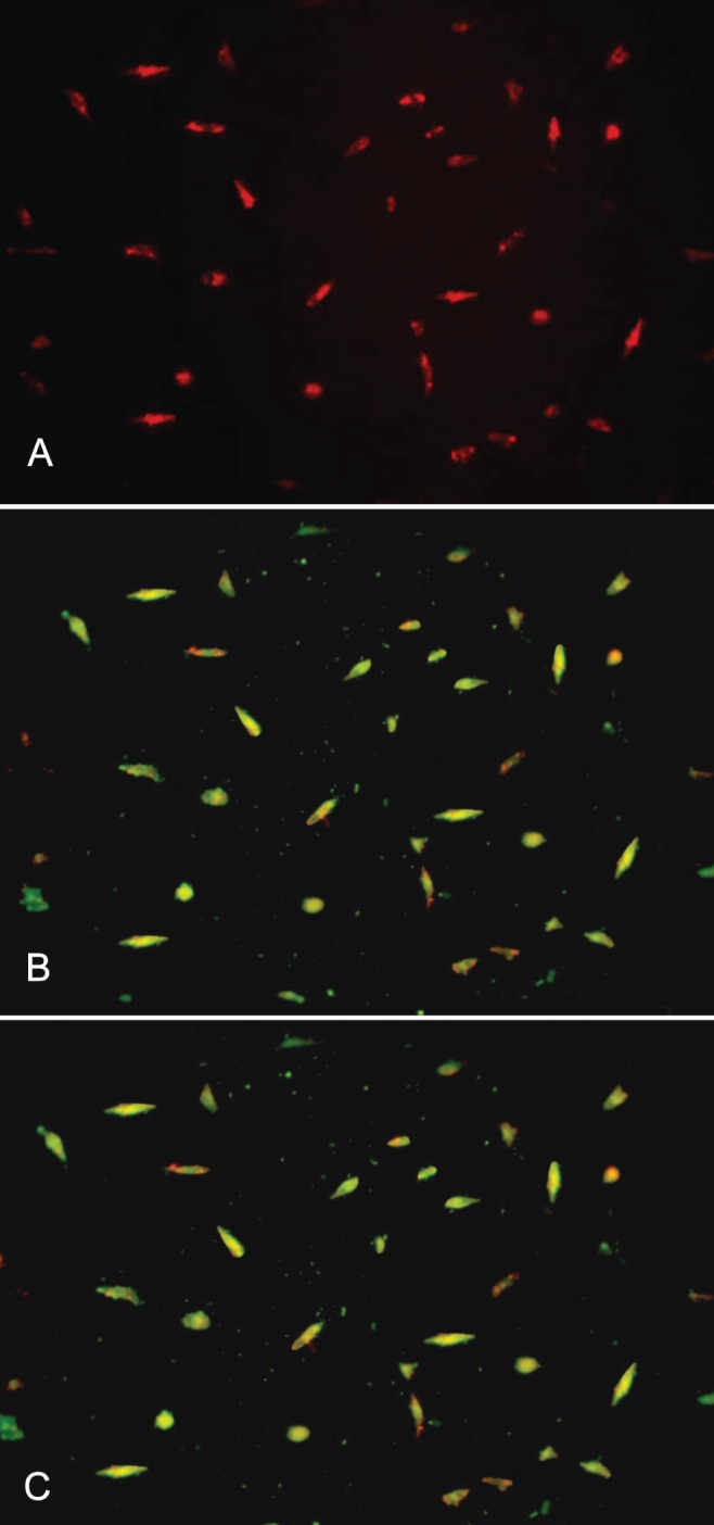
Figure 1).

Endothelial progenitor cells appear red after the uptake of dioctadecyl-tetramethylindocarbocyanine-labelled acetylated low-density lipoprotein (A), green after lectin binding (B), and yellow after double staining (C). Images were produced using inverted fluorescence microscopy (original magnification × 200)
