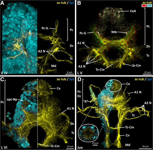
Figure 3.
Penilia avirostris, development of the visual system and the brain architecture (ventral view). A, C combined nuclei (cyan) and nervous system (yellow) image. B dorsally the compound eye anlagen (beige) and the remaining brain (yellow) depicted in 3D. D combined nuclei (cyan) and nervous system (yellow) image including a higher magnified image of a horizontal section of the compound eye. A antennula aesthetasc(s), A1 N antennula nerve, A2 N antenna nerve(s), ac-tub acetylated tubulin, Ce compound eye, CeA compound eye anlagen, Cn connective, Dc deutocerebrum, Lb-Cm labral commissure, Md mandibular neuromere, Ntb neurite bundle, opt-Np optical neuropil, Pc protocerebrum, Pc-lL protocerebral lateral lobes, Syt Sytox, Tc tritocerebrum, Tc-Cm tritocerebral commissure, asterisk ommatidia.

