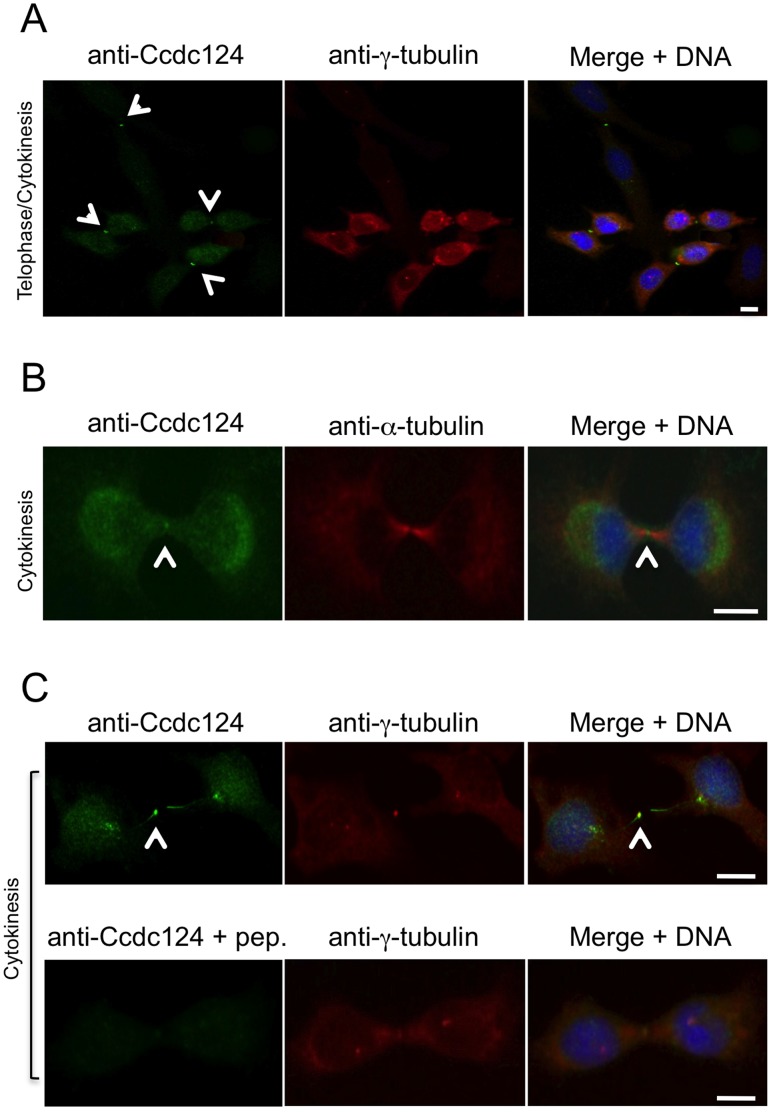
Figure 3. Ccdc124 accumulates at the midbody.
HeLa cells were synchronized as described in the legend of Fig. 2, and samples of cells were then stained with anti-Ccdc124 Ab together either with anti-γ-tubulin or with anti-α-tubulin Abs to monitor subcellular positions of centrosomes and the midbody. (A–B) At telophase and cytokinesis Ccdc124 is observed as puncta typically associated with the midbody positioned at the middle of intercellular bridge separating daughter cells, as detected in costainings with anti-γ-tubulin and anti-α-tubulin Abs, respectively. (C) Peptide competition assays were done by pre-incubating anti-N-ter-Ccdc124 antibody with the corresponding epitope peptide in 200-fold molar excess amounts. Signals generated by Ccdc124 localized at the midbody (shown with arrowhead) were lost in immunofluorescence assays where peptide pre-treated antibodies were used. Bars represent 10 µm.

