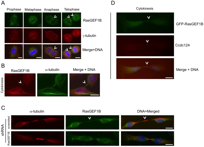
Figure 6. RasGEF1B and Ccdc124 colocalize at the midbody.
(A–B) Subcellular localizations of RasGEF1B proteins in synchronously dividing HeLa cells were detected with specific anti-RasGEF1B antibodies. Cell divisions were synchronized as described in the legend of Figure 2, above. Representative immunofluorescence microscopy images of HeLa cells costained with anti-RasGEF1B, and either anti-γ-tubulin (A) or α-tubulin (B) antibodies illustrating the position of MTOCs and the midbody at cytokinesis. Arrowheads show RasGEF1B detected at the midzone and the midbody. (C) Immunofluorescence signals observed at midbody were significantly decreased when endogenous RasGEF1B were depleted by transfections with specific shRNA vectors (Sh-C or Sh-D, Fig. S4) and representative micrographs were shown. (D) HeLa cells transiently transfected with GFP-RasGEF1B were fixed and stained using anti-Ccdc124 Abs. Arrowheads indicate midbody positions of GFP-RasGEF1B, Ccdc124, and their colocalizations at the midbody. Bars represent 10 µm.

