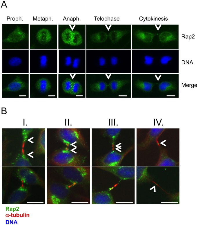
Figure 9. Active endogenous Rap2 relocates to midzone at anaphase, and to midbody during cytokinetic abscission.
(A) HeLa cells were arrested at G2/M phase by sequential double thymidine and nocodazole treatments as described in the legend of Figure 2, and they were classified according to phases of mitosis, and cytokinesis. Samples of cells were then stained with anti-Rap2 antibody, and with DAPI to visualize DNA. At anaphase Rap2 was detected at the midzone with staining characteristics reminiscent of endosomes, and at telophase/cytokinesis Rap2 was observed as puncta at the middle of the intercellular bridge, a position typically occupied by midbody associated factors. (B) Following synchronization of cells as above, 80 mins. after nocodazole was washed-off samples were taken with four consecutive intervals of 10 minutes (I, II, III, and IV), the last one (IV) corresponding to ∼120 minutes after the drug was removed, and dynamic positioning of Rap2 at the intercellular bridge in respect to α-tubulin was monitored. A time-dependent relocalization of Rap2 from peripheral flanking regions to the midbody was detected. Intercellular bridge localizations of Rap2 were concluded with observations from a sample of ∼50 cells in which over 75% showed similar positioning patterns. Two sets of representative micrographs were displayed. Bars represent 10 µm.

