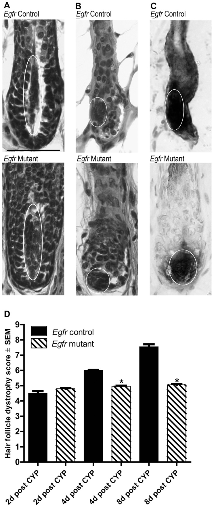
Figure 3. Cyclophosphamide-treated Egfr mutants displayed characteristics of early catagen with dermal papilla condensation 2d post administration.
A–B) Hematoxylin and eosin stained sections of vehicle treated controls (A top) and mutants (A bottom) displayed anagen dermal papilla with spindle shapes (ovals) surrounded by matrix keratinocytes. Control (B top) and mutant (B bottom) dermal papilla condensed to the proximal hair follicle by 2d following cyclophosphamide (ovals). Endogenous alkaline phosphatase confirmed dermal papilla position in controls (C top, oval) and mutants (C bottom, oval) at 2d. Scale bar indicates 50 µm. D) Hair follicle dystrophy score calculated following cyclophosphamide (CYP) as follows: dystrophic anagen = 1–3, dystrophic catagen = 4–6, dystrophic telogen = 7 and new anagen = 8. *Significant using Student’s t-test, where P≤0.05.

