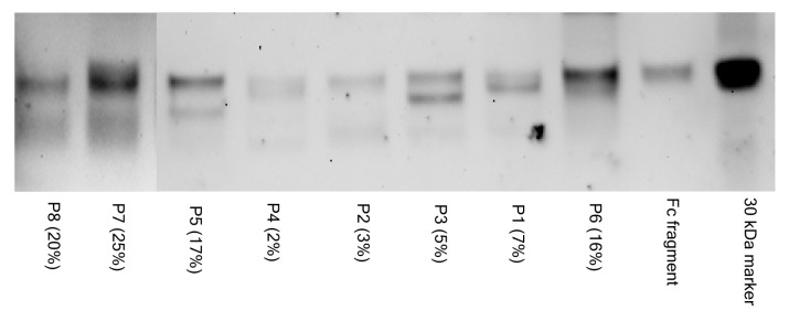
Figure 4. SNA lectin blot of Fc and Fab fragments from affinity purified anti-NY-ESO-1 IgGs. The percentage of sialic acid-containing (Sia+) IgGs observed in the course of the study is indicated in parentheses. The material analyzed by lectin blot consisted of pools of samples from the same patient that had been independently affinity purified. The image is a composite of two separate blots, as identified by the vertical line. The 30 KDa biotinylated marker and the commercially available Fc fragment (approximately 28 KDa) are shown for comparison. Lectin blot assays were done independently in duplicate instance, resulting in essentially identical findings. Each lane was loaded with approximately 1 μg of material.
