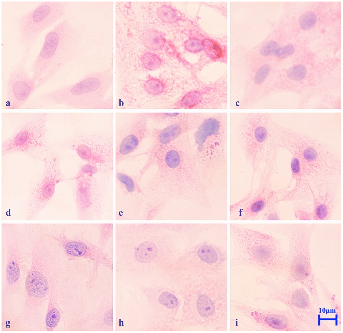
Figure 2. Pathological changes in primary rat myocardial cells exposed to heat stress in vitro at 42°C.
Myocardial cells were incubated at 37°C, heat stressed at 42°C for 10, 20, 40, 60, 120, 240, 360 and 480 min, stained with H&E and photographed using a Carl Zeiss optical microscope equipped with an imaging system (400×). Scale bar = 10 µm. a. Control cells (37°C). b. After 10 min of heat stress, acute granular degeneration was observed in the cytoplasm compared to control cells. c. After 20 min of heat stress, the cytoplasm of the swelling myocardial cells was obviously cloudy. d. After 40 min, numerous red granules were observed in the cytoplasm. e. After 60 min of heat stress, the nuclei were observed to be markedly basophilic and the cell sizes were enlarged. f. After 120 min of heat stress, karyopyknosis was observed. g. After 240 min, markedly basophilic nuclei and intracellular vacuoles were observed in the heat stressed myocardial cells. h. After 360 min, the number of intracellular granules in the cytoplasm of swollen myocardial cells decreased. i. After 480 min of heat stress, the cells remained enlarged and degeneration was evident compared with the control group.

