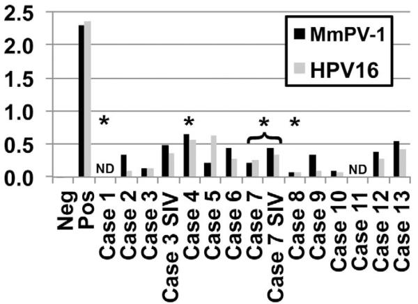
Figure 5.
Papillomavirus serologic screening by ELISA of the 13 cases examined within this study. Titers against both rhesus papillomavirus 1 (MmPV-1, black bars) and human papillomavirus 16 (HPV 16, gray bars) were low in all cases and were comparable to controls (Cases 9–13). Paired pre- and postinfection sera from experimentally infected SIV+ animals (Cases 3 and 7) are shown. Asterisks indicate animals with positive papillomavirus E6 antigen staining by immunohistochemistry. ND: not determined; serum unavailable for analysis. Controls include a nonspecific mouse IgG (negative) and an HPV 16 L1 anti-capsid antibody (positive).

