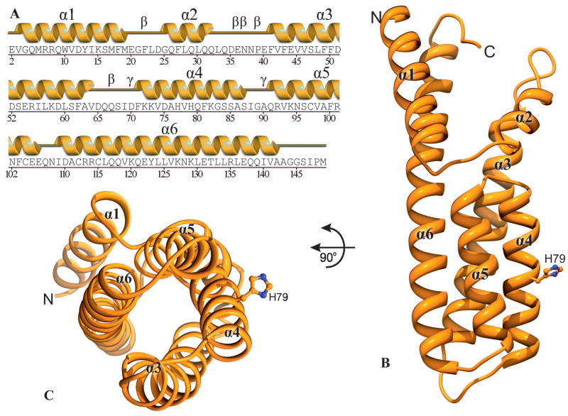
Fig. 2.
Overall structure of MtHPt1. A Secondary structure elements (helices α1- α6) corresponding to the amino acid sequence. Five β and two γ turns are also labeled. B A ribbon diagram illustrating the six α-helices and the phosphorylation site His79. Four of the helices (α3-α6) form a helix bundle. C View down the axis of the four-helix bundle. The His79 side chain of the active site is highly exposed to the solvent.

