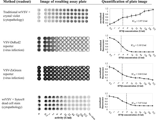
Figure 2.
Comparison of live/dead cell stains and fluorescent-protein expressing assay virus. A549 cells were incubated under serial 2-fold dilutions of recombinant human IFNβ for 24 hours, then infected with either wild-type or recombinant VSV as indicated at a multiplicity of 5 pfu/cell. After 24 hours of infection, assay plates were stained, imaged, and quantified as discussed in the Methods section. Positive signal indicates cell survival for the crystal violet assay, virus replication for the fluorescent virus assays, and cell death in the Sytox assay. Positive control wells are cells untreated by antivirals and infected. Negative control wells are untreated, uninfected cells.

