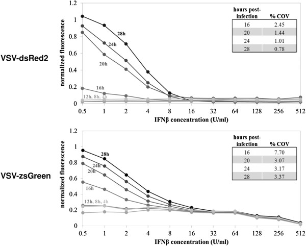
Figure 3.
Assay time-course development. Recombinant VSV antiviral activity assays were imaged on a fluorescent biomolecular imager every four hours post-infection to monitor fluorescent signal development indicating viral replication. Mean fluorescence values were extracted from plate images, normalized to positive and negative controls and plotted. Darker markers indicate longer development times. IC50 values were calculated as described, and the coefficient of variance was calculated between four replicates at each time point.

